
|
 |
Canine Hip Dysplasia (CHD) is a developmental disorder of the hip that
begins with joint laxity and progresses to arthritis over a period of several months
to years. It is one of the most common skeletal diseases seen by veterinarians.
The condition is very common in large breed dogs, but can be seen in any breed.
Multiple genes are involved in the inheritance of hip dysplasia, and
many other factors influence its development, including body type, size, growth
rate, and nutrition. overfeeding, and dietary supplementation for maximal growth
has been shown to increase the incidence of hip dysplasia in young, growing, large
breed dogs. Conversely the development of hip dysplasia can be delayed, and its
severity diminished when the growth rate of pups is restricted.
|
|
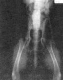
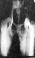
Normal Hips
|
|
|
 |
|
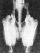
Severe Hip Dysplasia
|
|
Diagnosis |
|
|
|
The diagnosis of hip dysplasia is based on history, physical examination,
and radiographic evaluation. A typical history may include any or all of the following:
- difficulty or stiffness upon rising
- rising using front legs only and dragging rear
- "bunny hopping"'gait
- short stride in rear legs
- reluctance to exercise or climb stairs
- rear limb lameness
- soreness in hips
- waddling rear limb gait
The clinical signs commonly begin between five to eight months of age
or after skeletal maturity. Some dogs don't have noticeable problems until eight
to ten years of age or older. The onset of clinical signs may appear sudden or gradual.
This variability is due to the individual severity of the disease as well as pain
tolerance of the pet.
Most dogs with CHD are most painful when the hips are extended by pulling
the rear legs back behind the body. Palpation of the hips usually reveals joint
laxity, although anesthesia may be required to detect it in some cases.
Radiographs are necessary to confirm the diagnosis and evaluate the severity
of CHD. In young dogs or in very mild cases, joint laxity may be the only detectable
abnormality. Later in the disease arthritic changes are seen. The standard radiographic
position is with the dog lying on its back with both rear legs pulled straight back
and parallel to each other. Most dogs with CHD are too painful to tolerate this
position awake, so sedation or anesthesia is usually necessary. Proper radiographic
positioning is very important to accurately evaluate the hips and to determine the
best treatment.
The
Orthopedic Foundation of America (OFA)
has been the standard for
certification of dog's hips as being free of CHD. The radiograph is taken after
the dog is 2 years of age and requires the hip extended position. Unfortunately,
our progress has been disappointing in reducing the frequency of CHD using OFA alone.
Recently the PennHIP program has emerged as a new scientific method for the early
diagnosis of CHD. It measures the passive hip joint laxity or "looseness" of the
hip ball in the hip socket under sedation or anesthesia. PennHIP is more reliable
and has the advantage of being accurate on puppies as young as 16 weeks of age.
Dr. Henney is trained and certified to perform the PennHIP procedure.
|
|
Treatment |
|
|
|
Medical and/or surgical treatment may be recommended for CHD, depending
on the individual circumstances. Medical management usually consists of exercise
restriction, body weight management, and symptomatic pain management with analgesics
and anti-inflammatory drugs. A non-weight bearing activity like swimming is the
preferred type of exercise since it places minimal stress on the joints.
Several surgical options are now available for treating the various stages
of hip dysplasia. They include the triple pelvic osteotomy, femoral head and neck
excision, and total hip replacement.
|
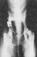
Triple Pelvic Osteotomy |
 |
|
The ideal candidate for this procedure is a young dog (5 months to I
year of age) with clinical signs of CHD, including hip joint pain and laxity, but
little or no radiographic signs of arthritis. In this procedure the acetabulum (hip
socket) is rotated to a more normal position in relation to the femoral head. This
seats the head more deeply into the socket. The pelvis is then stabilized in this
new position with a specially designed pelvic osteotomy plate and bone screws. The
procedure eliminates the joint laxity and corrects the abnormal conformation of
the socket. Thus, the hip is able to develop more normally without developing arthritis.
overall, with proper case selection, it appears that approximately 90% of dogs achieve
good to excellent results and return to athletic endeavors. Owners have been uniformly
very pleased with the results and have had no reluctance recommending the procedure
to others. If a TPO is performed on the opposite leg, it is performed 3-6 weeks
later. |
|
|
|
|
Femoral Head and Neck Excision |
 |
|
When arthritis develops in a dog with hip dysplasia the damage is irreversible,
so a triple pelvic osteotomy will not restore normal hip function or eliminate the
pain. instead, a salvage procedure is needed that removes the source of pain. Two
such procedures are the femoral head and neck excision and the total hip replacement.
The femoral head and neck excision is the most commonly performed procedure. It
involves removing the femoral head, which is the ball portion of the joint. The
body's reaction is to form a false joint, which along with the surrounding pelvic
muscles minics joint function. Although this does not return the hip to normal,
it significantly relieves the pain of arthritis. making the leg more functional
and comfortable. Thus, the dog usually becomes more active. The advantage of this
procedure is that it is relatively simple, has few complications, can be performed
at any age, is relatively inexpensive and has good results in the majority of dogs.
Best results are obtained in small and medium sized dogs. |
|
|
|
|
Total Hip Replacement |
 |
|
This procedure involves removal of both the ball and socket portions
of the hip joint and replacing them with artificial implants. It is currently the
best available treatment for severe hip dysplasia in large, mature dogs. Currently,
the success rate is reported to be over 95%, and dogs with successful implants are
able to perform almost any task performed by dogs with normal hips. Therefore, it
is the treatment of choice for dogs used for working or sporting activities or when
optimal hip function is desired.
Candidates for total hip replacement are typically large breed dogs with
irreparable disease or injury to the hip joint. The dogs must be fully grown ( minimum
9 months of age), over 40lbs, free of any infections, debilitating conditions, or
neurologic disorders. Their health is fully evaluated before surgery to minimize
the risk of complications. Despite extensive precautions, complications are reported
in approximately 10% of the cases. Many are correctable, but some serious complications
may require removal of the implants. Proper home care after surgery is very important
to the success of the procedure. Strict confinement and close supervision is required
for the first eight weeks after surgery. 80% of cases require surgery on only one
side. Ultimately most dogs regain full, pain free function after total hip replacement,
and live normal active lives.
|
|
|
|